SOFIA

[COUNTER_NUMBER id=446]

SOFIA 3D Breast Ultrasound is making breast cancer imaging more accurate, efficient, and comfortable. The whole breast can be scanned in 30 seconds.  Appointments can be completed in 10 minutes. and radiologist can read the resulting stores in 2 minutes.

3D Breast Ultrasound  in seconds

The SOFIA 3D Breast Ultrasound was developed with dense breast women  in mind.

Throughout the scan the patient remains covered, lying comfortably on the memory foam bed, with no compression of her breasts.

SOFIA can image an entire breast in 30 seconds.

The 3D images make it much easier for a radiologist to view the patient's breast tissue on a SOFIA Advanced Workstation.

All patients deserve the best most reliable care.

Unrivaled Efficiency

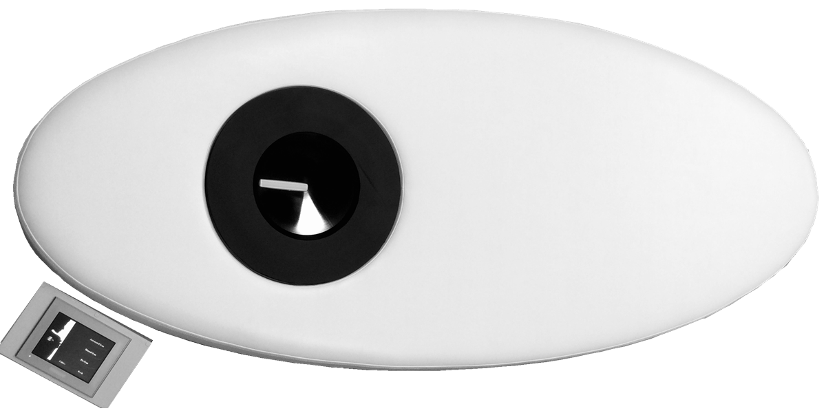
  • Large 200mm field of view, state of the art automation, and prone patient positioning
  • Enabling image acquisition of the entire breast in a 30 second scan
  • Resulting in a single 3D volume per Breast
This is an image of a bilateral breast 3D Automated Breast Ultrasound (ABUS) exam of a female heterogeneously dense breast (BI-RADS C) .  Both the left an right breast are displayed are displayed on the monitors for rapid comparison.  The axial  and coronal slices of each breast are displayed.  The coronal image aids in determining the location of structures and the relative location of structures to one another.

The multi-planar reconstruction (MPR) enables the radiologist to view any structure from any perspective.  Thereby eliminating the need for call backs to obtain additional images from different perspectives.

The radiologist has the capability to window level, zoom-in zoom-out, pan, measure linearly, measure volumes, rotate 3D volume, rotate plane, adjust slice thickness, thin MIPS, synchronize review, compare studies for breast cancer therapeutic monitoring.

The SOFIA 3D Automated Breast ultrasound (ABUS) exam is capable of penetrating through Extremely Dense Breast Tissue (BI-RADS D) and   Heterogeneously Dense Breast Tissue (BI-RADS C) enabling visualization to the pectorals muscle.

Rapid Case Review

  • 2 minute review per bilateral case
  • Intuitive user interface and functionality
  • DICOM compatible data sets
This is a 3D Automated Breast Ultrasound (ABUS)  image of a female heterogeneously dense breast (BI-RADS C)  that contains multiple cysts of varying sizes. Some have irregular margins.  

The upper image is an axial slice of the breast shows the breast tissue and structures between 10 o'clock and 4 o'clock.

The lower image is a coronal slice of the breast that shows the location of the various cystic structure and there relative location to each other.

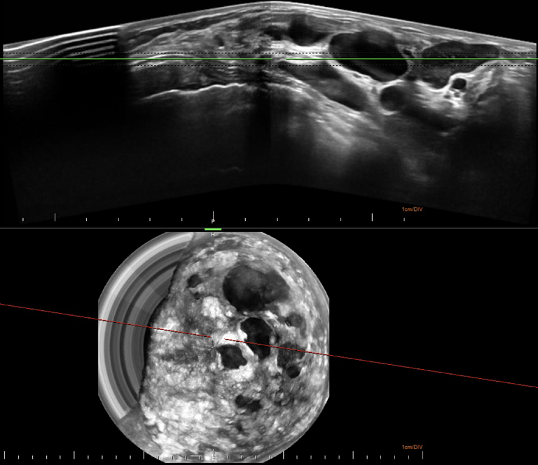
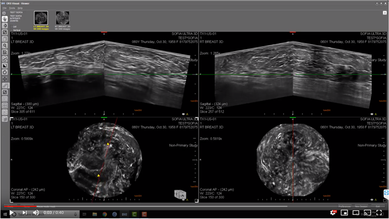
Excellent Image Quality

  • 3D whole breast diagnostic quality images
  • Correlates with manual B-mode images
  • Multiple viewing angles for any anatomical feature within the breast
This is an image of a women comfortably lying on the SOFIA examination table in  the prone position during the 3D Automated Breast Ultrasound (ABUS) Exam of her breast.

We understand how important privacy is to some patients.  Therefore during the examination the patient can remain covered during the 3D Automated Breast Ultrasound (ABUS) exam.  

SOFIA was developed based on patient input instead of attempting to apply technology to patients.  

Potential patients  advised us that they were looking for a way to routinely have their breast checked for breast cancer that was:
comfortable, private, natural, painless, holistic, compression-less, ion radiation-free, rapid, accurate,  efficient, eliminates callbacks, 3D, comprehensive,

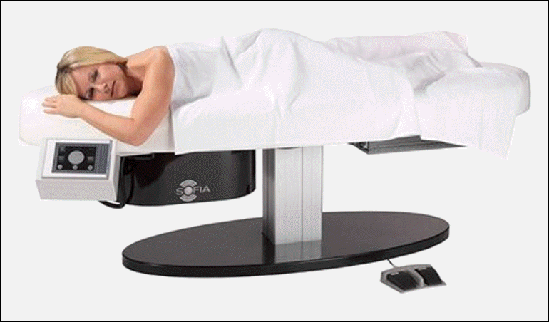
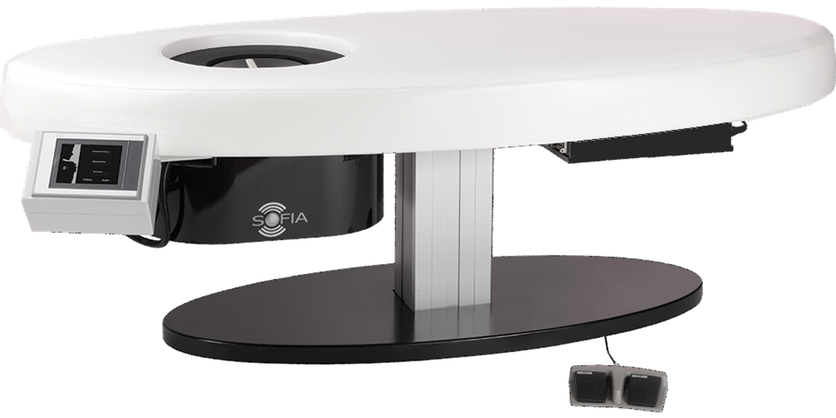
Unsurpassed Patient Comfort

  • Prone Patient Positioning on a memory foam examination table
  • Warm acoustic lotion provides a comfortable interface between the transducer and the patient’s breast
  • Rapid non-compression exam
Women with dense breast are 4 to 5 times more likely to develop breast cancer.

Throughout her life a woman has 1 in 8 chance of developing breast cancer.

Mammograms are essential to detect breast cancer early so it can be treated. However, 40% of women have dense breasts making cancer more difficult to detect.

A large multi-center medical trail found that 55% more cases of cancer were detected in women with dense breast with the addition of ultrasound.

The SOFIA 3D breast ultrasound was developed for women with dense breast tissue.

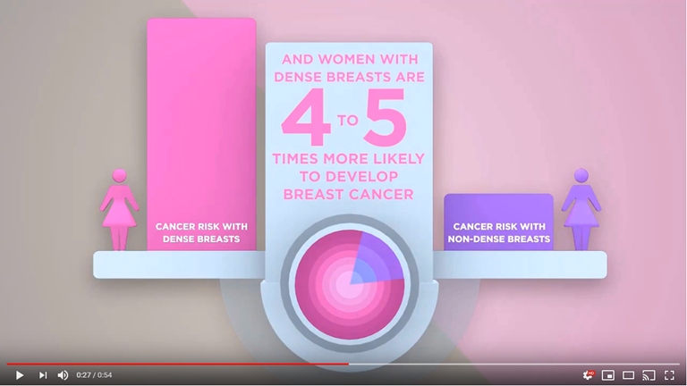
Addressing the needs of Dense Breast Patients

  • 40% of patients who have a mammogram have dense breast tissue
  • Increased Breast Density reduces the sensitivity and specificity of mammography
  • Supplemental imaging with whole breast ultrasound can detect cancers not seen on mammography in this patient population
SOFIA was developed to seamlessly integrate in today's modern healthcare information technology architectures composed of: Picture Archive and Communications Systems (PACS), Radiology Information Systems (RIS), Vendor-Neutral Archive (VNA), Electronic Health record (EHR), Electronic Medical Record (EMR), Virtual Private Network (VPN), Digital Imaging and Communications in Medicine (DICOM), Multiplanar Reconstruction (MPR), Health Insurance Portability and Accountability Act (HIPAA), Health Level 7 (HL7), ISO 27001, ISO 27799, Health Information Trust Alliance (HITRUST CSF), and HTML 5 Web Browser.

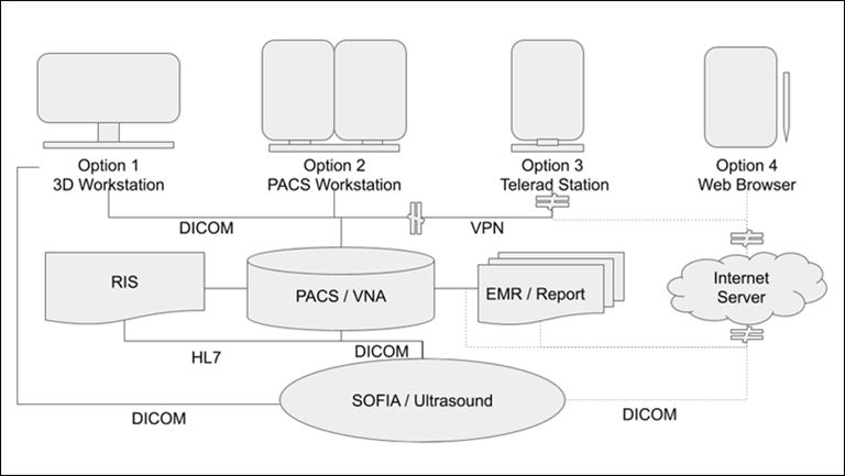
SOFIA Review Architecture

  • User-definable hanging protocols based on radiologist preferences
  • Flexible review architecture (dedicated workstation, cloud based interface, multi-site, multi-user configurations)
  • Leverages existing IT infrastructure investment
  • Built on standards HL7, DICOM, VPN, Security practices enabling integration with PACS, RIS, and EMR systems.

To learn more about SOFIA 3D Breast Ultrasound

SOFIA™ by iVu Imaging Corporation
935 South Kimball Avenue, Suite 166, Southlake, TX 76092 USA
Copyright © 2019 iVu Imaging Corporation