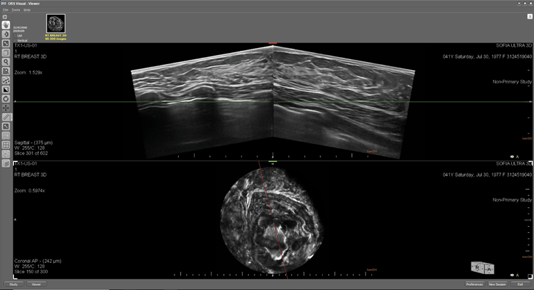
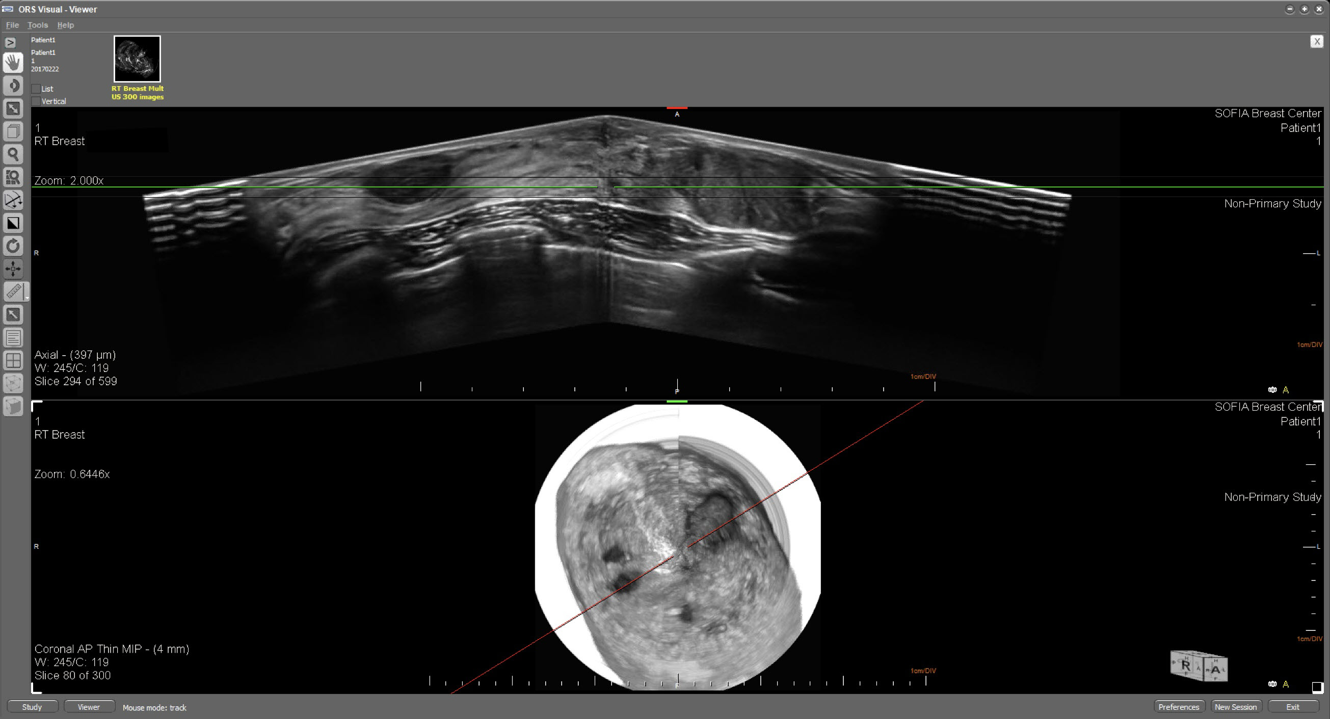
Normal Dense Breast

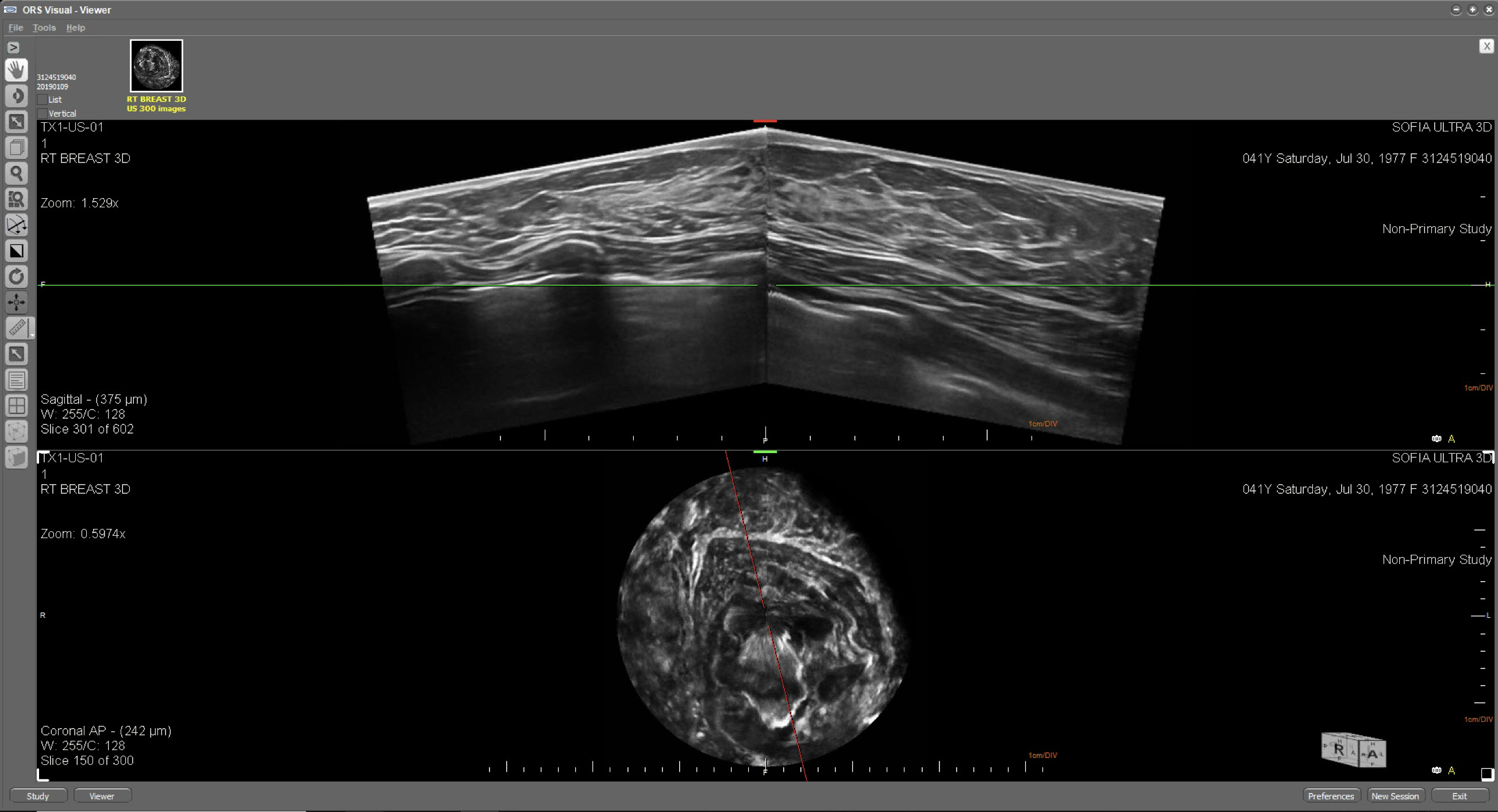
Normal Breast Implant
Simple Cyst
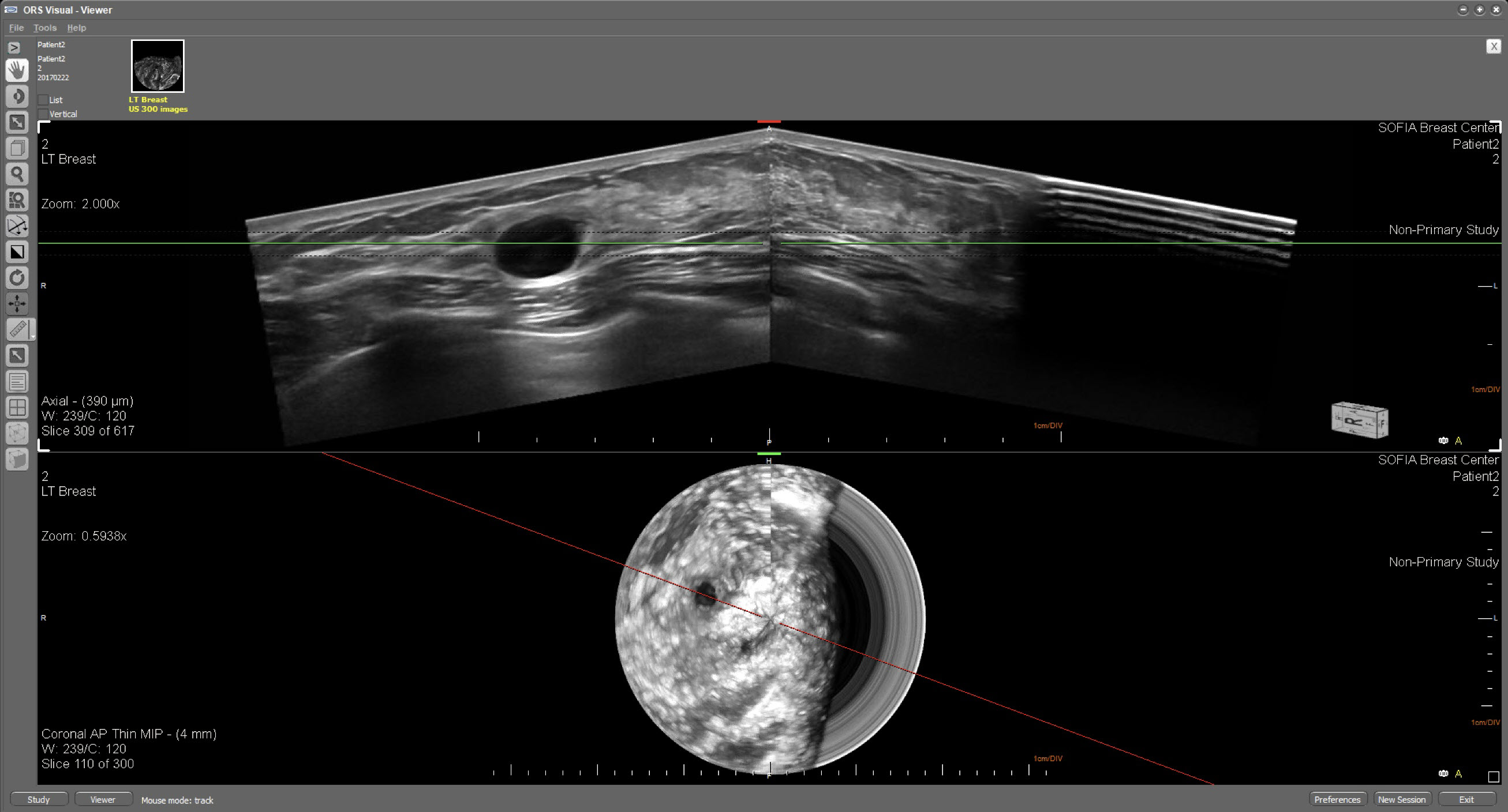

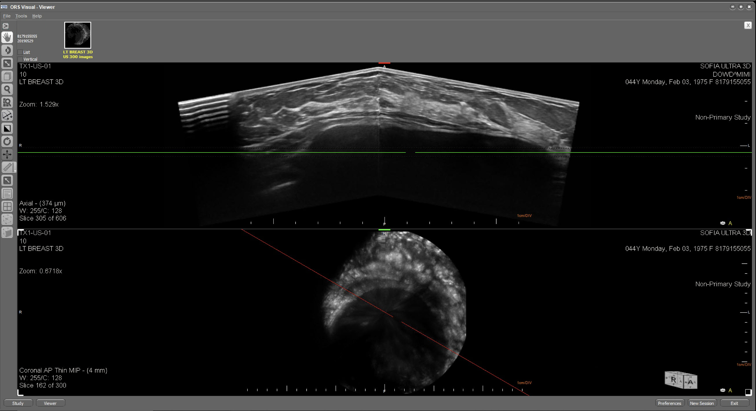
Multiple Cysts
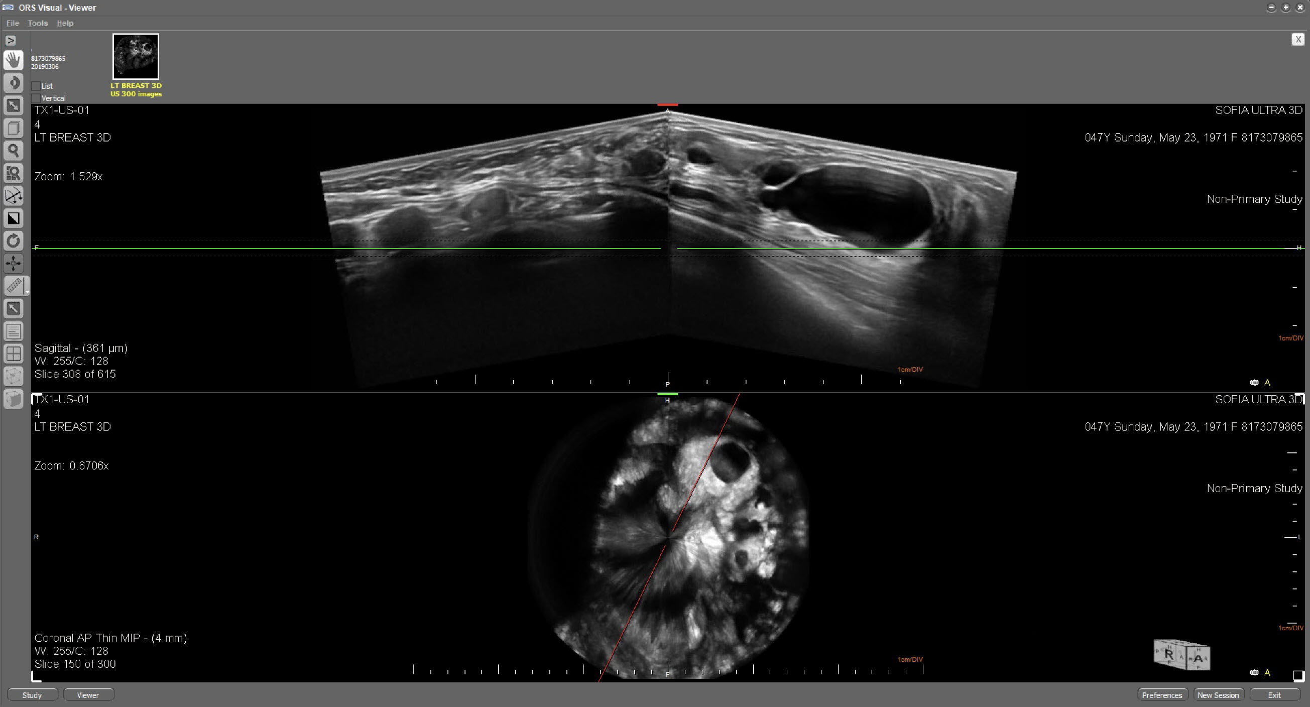
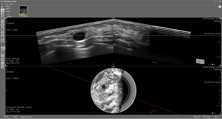
Complicated Cyst
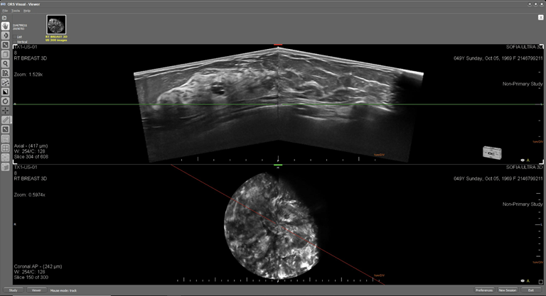
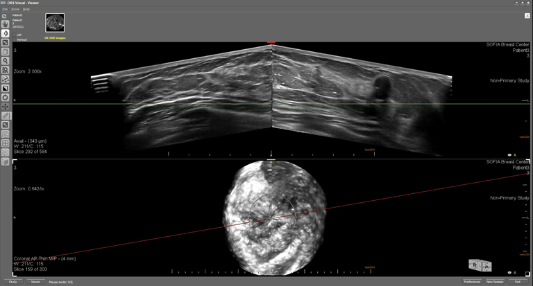
Multiple Fibroadenomas
Solid Nodule with Microcalcifications
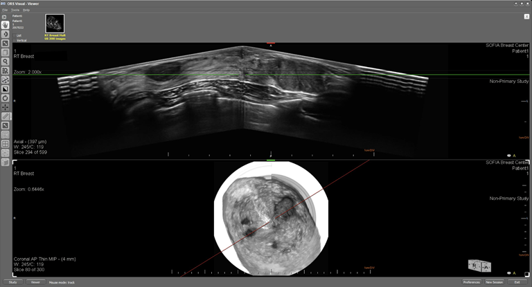
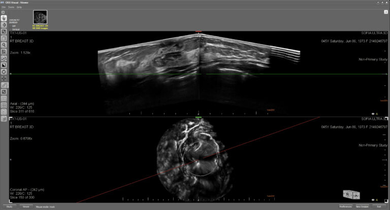
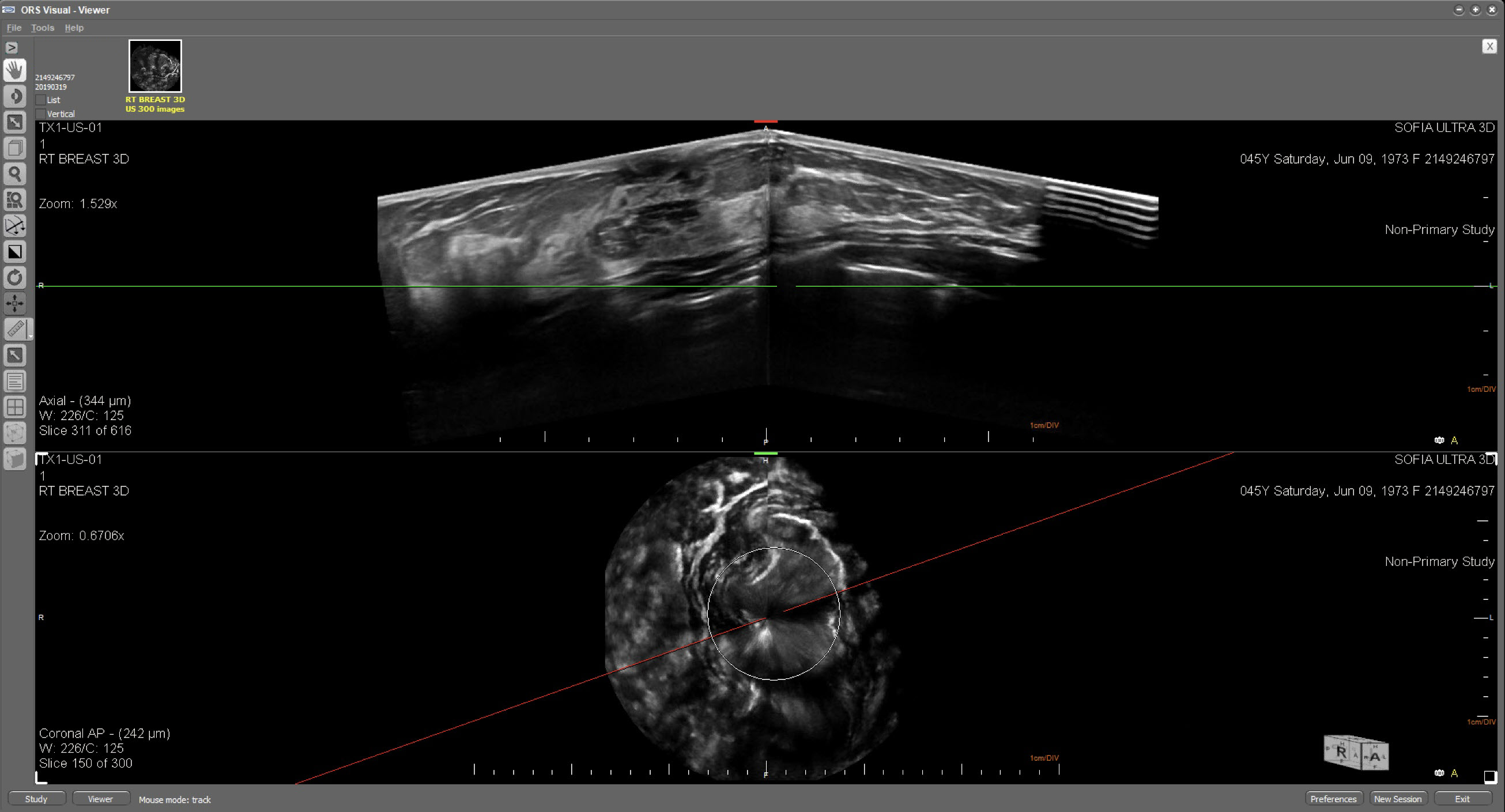
Biopsy Proven Cancer
To learn more about SOFIA 3D Breast Ultrasound

![]()
SOFIA™ by iVu Imaging Corporation
935 South Kimball Avenue, Suite 166, Southlake, TX 76092 USA
Copyright © 2019 iVu Imaging Corporation