Discover a Better Way to Screen for Breast Cancer

Automated 3D Breast Ultrasound with SOFIA®

Reimagine In-Office Breast Cancer Imaging

SOFIA is an innovative, FDA-approved ultrasound screening technology specifically designed for early detection of cancer in dense breast patients.

Effective: Up to 2x more accurate diagnosis results for patients with dense breast tissue1
Efficient: Cost-effective and profitable in-office procedure performed by your existing team
Comfortable: A better patient experience, without compression, injections, or radiation

Discover if SOFIA is right for your practice by scheduling a virtual or in-person demo - just complete the form to get started.

Schedule a Demo

Discover if SOFIA 3D breast ultrasound is right for your OBGYN practice by scheduling a demo.

Thank you! Your submission has been received!
Oops! Something went wrong while submitting the form.

Revolutionizing 3D Breast Ultrasound with SOFIA

SOFIA combines speed, accuracy, and patient comfort in one seamless 3D breast ultrasound system for earlier detection of cancer for all patients, including those with mammographically dense breasts. Discover why more OBGYN practices are expanding their in-office breast imaging capabilities with SOFIA:

SCHEDULE DEMO

Is Mammography Putting Your Current Patients at Risk?

Dense breast tissue obscures early stage cancer on a mammogram, leading to a 50% missed diagnosis rate in comparison to 3D ABUS.2

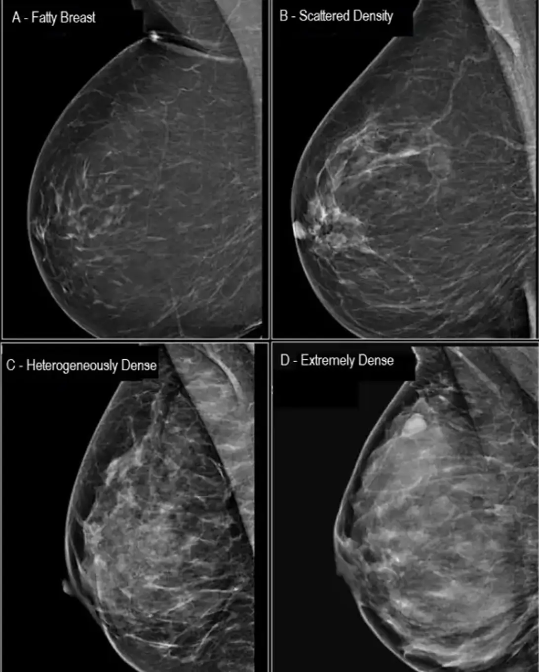
Research shows that women with mammographically dense breasts (about 40% of the patient population3) are at higher risk of developing breast cancer - up to 6x more likely than other patients. While mammography is well-known as the first line of defense for breast health screening, it’s significantly less sensitive in patients with dense breasts.
EXPLORE OPTIONS

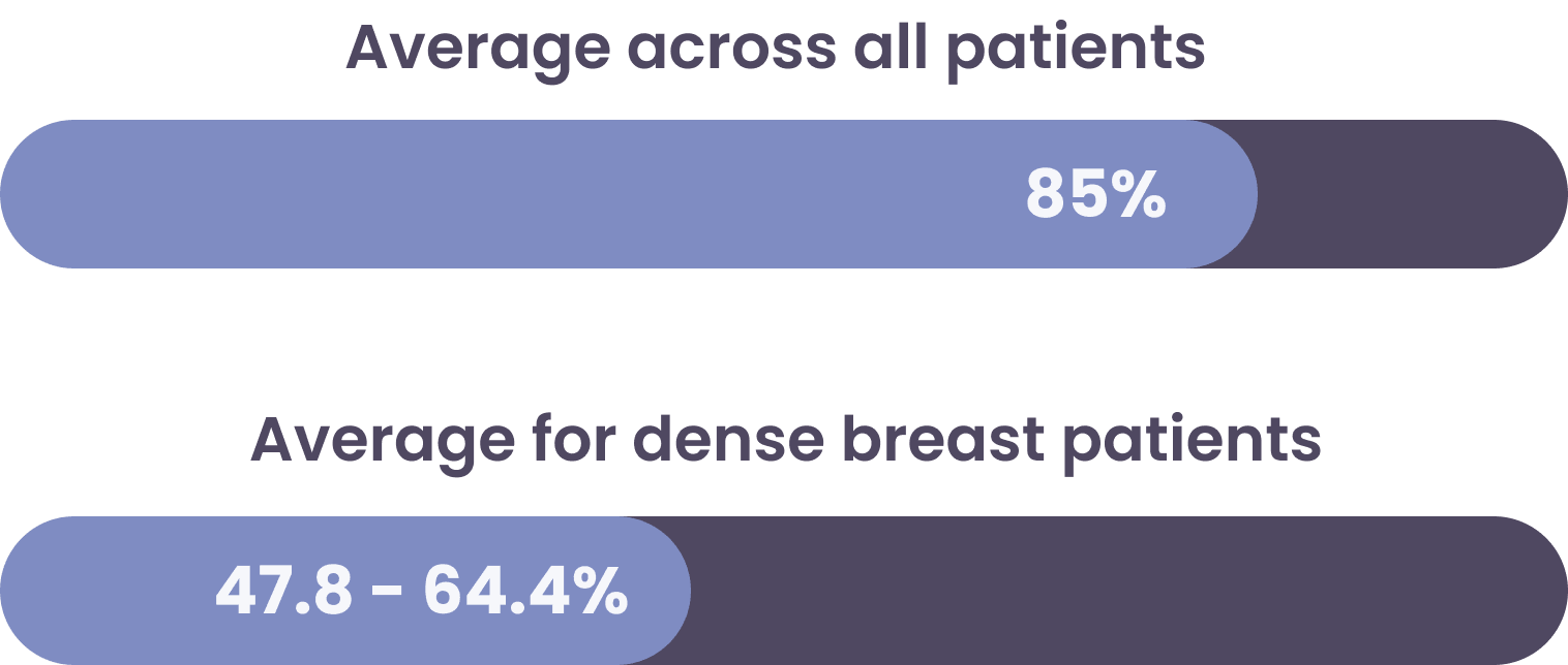
Mammography Sensitivity Comparison4:

Dense breast tissue and cancer both present as white on mammograms, hiding small or early-stage cancers. As a result, mammography’s sensitivity and ability to detect cancer is significantly reduced for dense breast patients

Early cancer detection rates are significantly increased with supplemental 3D Ultrasound like SOFIA. Find out how your patients and practice can benefit:

EXPLORE OPTIONS
CLINICAL INSIGHTS

Comparing Mammography Alone vs. 3D Breast Ultrasound for Cancer Detection

“Integrating full-field digital mammography (FFDM) with 3D prone ABUS in women with high breast density increases and improves breast cancer detection rates in a significant manner, including small and invasive cancers, and it has a tolerable impact on recall rate. Moreover, 3D prone ABUS performance results are comparable with the performance results of the supine 3D ABUS system.1

Journal of Personalized Medicine, 2021
Second-Generation 3D Automated Breast Ultrasonography (Prone ABUS) for Dense Breast Cancer Screening Integrated to Mammography: Effectiveness, Performance and Detection Rates

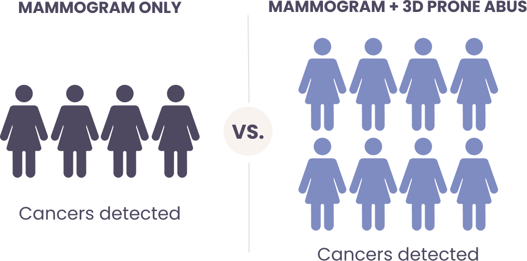
Cancer Detection Rate:
Mammography vs. ABUS


In a 2021 clinical study, 3D breast ultrasound identified cancer at twice the rate of mammogram only for dense breast patients.1
How It Works:

SOFIA 3D ABUS

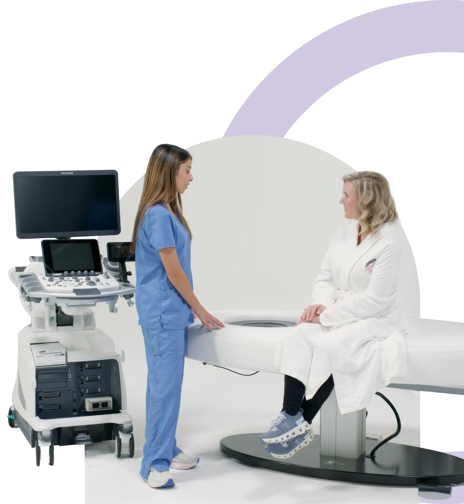
SOFIA’s unique scanning cone is recessed into its comfortable scan bed and houses Hitachi’s extra-long linear probe. The patient lies in the prone position placing her breast with the nipple in the center. In just a single 30-second radial sweep, a 3D volume dataset is collected of the entire breast.
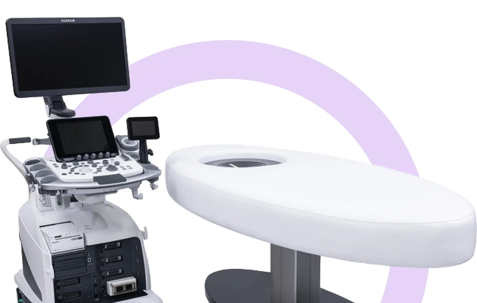
Thirty-second, per-breast scan time allows the scheduling of patients as often as every ten minutes
An intuitive touch-screen interface automates the scanning process for in-office imaging teams
The procedure requires only a thin layer of ultrasound lotion – no additional disposables required
Results quickly readable by local radiologist or breast teleradiologist

Frequently asked questions

Can SOFIA be installed in one of my existing exam rooms?

Yes, SOFIA typically can be installed in an exam room without any modifications, and the room can still be used for regular exams.

How long does it take to install a SOFIA system in my exam room?

SOFIA can be installed in the morning, and that afternoon, your fully trained staff can be seeing patients and performing exams.

Is specialized training required to operate the SOFIA system?

Most facilities have their existing mammographers or sonographers perform the SOFIA Breast exams.

Who reads the SOFIA breast ultrasound exams?

The SOFIA Breast ultrasound exams can be read by your local radiologist, or we can connect you with a fellowship-trained breast radiologist.

I thought 3D Mammography was supposed to be the answer for my dense breast patients.

The sensitivity and specificity of mammography are reduced in dense breast patients, which can lead to missed cancers.

How long does a typical SOFIA breast ultrasound exam take, including pre/post-exam time?

Most facilities schedule SOFIA exams for a 15-20 minute appointment slot. This includes time for patient Q/A, data entry, positioning, and prep for the next exam.

Does healthcare insurance pay for SOFIA breast ultrasounds? Do copays and deductibles apply?

Over 35 states have laws specifically related to healthcare coverage for SOFIA breast ultrasound exams. In addition, some states have passed additional legislation eliminating co-pays and deductibles, thereby removing patient out-of-pocket costs. For state-by-state details, visit the interactive insurance map on DenseBreast-info.org.

What disposables are required to perform a SOFIA breast ultrasound exam?

Just a small amount of warm ultrasound lotion and sanitizing wipes for between patients. Some facilities also enhance the patient's experience by providing a warm face towel and spa robe.

Discover how SOFIA can transform breast cancer imaging for your practice.

SCHEDULE DEMO
  1.  Journal of Personalized Medicine, 2021: Second-Generation 3D Automated Breast Ultrasonography (Prone ABUS) for Dense Breast Cancer Screening Integrated to Mammography: Effectiveness, Performance and Detection Rates
  2. Comparison of the Performance of Screening Mammography, Physical Examination, and Breast US and Evaluation of Factors that Influence Them: An Analysis of 27,825 Patient Evaluations Thomas M. Kolb, Jacob Lichy, Jeffrey H.
  3. Pisano et al. NEJM 2005; 353: 1773
  4. https://www.breastcancer.org/research-news/breast-density-affects-contralateral-bc-risk